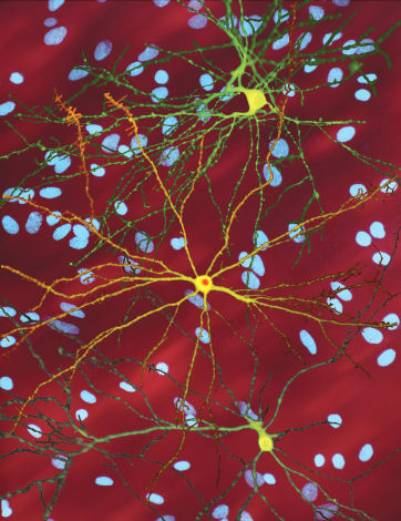
File:Neuron with mHtt inclusion.jpg
Neuron_with_mHtt_inclusion.jpg (362 × 470像素,文件大小:60 KB,MIME类型:image/jpeg)
文件历史
点击某个日期/时间查看对应时刻的文件。
| 日期/时间 | 缩略图 | 大小 | 用户 | 备注 | |
|---|---|---|---|---|---|
| 当前 | 2009年3月14日 (六) 23:39 |  | 362 × 470(60 KB) | Leevanjackson | {{Information |Description={{en|1=single striatal neurons (yellow) transfected with nuclear inclusion (orange) mHtt, other neurons in background (blue), from press release http://www.ninds.nih.gov/news_and_events/press_releases/pressrelease_huntington%27s |
文件用途
以下页面使用本文件:
全域文件用途
以下其他wiki使用此文件:
- af.wiki.x.io上的用途
- ar.wiki.x.io上的用途
- azb.wiki.x.io上的用途
- az.wiki.x.io上的用途
- bg.wiki.x.io上的用途
- bs.wiki.x.io上的用途
- ca.wiki.x.io上的用途
- da.wiki.x.io上的用途
- el.wiki.x.io上的用途
- en.wiki.x.io上的用途
- en.wikiversity.org上的用途
- eu.wiki.x.io上的用途
- fa.wiki.x.io上的用途
- fr.wiki.x.io上的用途
- ga.wiki.x.io上的用途
- he.wiki.x.io上的用途
- hi.wiki.x.io上的用途
- hu.wiki.x.io上的用途
- hy.wiki.x.io上的用途
- id.wiki.x.io上的用途
- it.wiki.x.io上的用途
- it.wikibooks.org上的用途
- ja.wiki.x.io上的用途
- jv.wiki.x.io上的用途
- kn.wiki.x.io上的用途
- lmo.wiki.x.io上的用途
- mk.wiki.x.io上的用途
- ms.wiki.x.io上的用途
- no.wiki.x.io上的用途
- or.wiki.x.io上的用途
- pl.wiki.x.io上的用途
- pt.wiki.x.io上的用途
- ru.wiki.x.io上的用途
- sco.wiki.x.io上的用途
- sh.wiki.x.io上的用途
- simple.wiki.x.io上的用途
- si.wiki.x.io上的用途
- sl.wiki.x.io上的用途
- sq.wiki.x.io上的用途
- sr.wiki.x.io上的用途
- sv.wiki.x.io上的用途
- te.wiki.x.io上的用途
查看此文件的更多全域用途。

