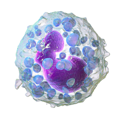
嗜鹼性球
嗜碱性粒细胞或嗜碱性球(英語:Basophil、Basophilic granulocyte)是最少見的一種粒細胞,約佔循環系統中白血球的0.5%至1%[1]。然而,嗜鹼性球是粒細胞中最大的一類。嗜鹼性球是許多免疫反應(特別是過敏反應)中促進發炎反應的細胞,與過敏性休克、氣喘、異位性皮膚炎、過敏性鼻炎等過敏性疾病有關[2]。
| 嗜鹼性球 | |
|---|---|
 嗜鹼性球的電腦模型 | |
 經染色的血液樣本,在嗜鹼性球周圍的紅色細胞為紅血球 | |
| 基本信息 | |
| 系統 | 免疫系統 |
| 功能 | Granulocyte |
| 标识字符 | |
| MeSH | D001491 |
| TH | H2.00.04.1.02022 |
| FMA | FMA:62862 |
| 《显微解剖学术语》 [在维基数据上编辑] | |
嗜鹼性球於1879年由德國生物學家保罗·埃尔利希發現,而他於前一年(1878年)亦首次描述了肥大細胞[3]。
嗜鹼性球的細胞質中有許多大的顆粒,在顯微鏡下容易與細胞核混淆,但當尚未染色時細胞核就清晰可見且通常分成兩葉[4]。肥大細胞與嗜鹼性球有許多相似之處,如兩種細胞都存有組織胺,一種會在特殊情形下被刺激釋放的化學物質。如同大多數的粒細胞,嗜鹼性球在需要時可由血液進入組織中。
嗜鹼性球的命名來自於其可以被鹼性的染劑染上色的特性。
功能
编辑嗜鹼性球參與了許多形式的發炎反應,尤其是造成過敏的種類。嗜鹼性球中含有稱為肝素的抗凝血劑,可避免血液太快凝集,也含有血管擴張劑組織胺,可促進血液流至組織中,嗜鹼性球在受寄生蟲感染的部位會異常增高,如同嗜酸性球,嗜鹼性球在對抗寄生蟲感染與過敏反應都扮演重要的角色,大量出現於發生過敏的組織中[5]。嗜鹼性球的細胞膜表面有可與IgE結合的蛋白質受體,IgE是一種參與對抗寄生蟲感染與過敏反應的抗體,結合在嗜鹼性球上的IgE可對環境中的物質產生選擇性的過敏反應,如花粉粒或某些寄生蟲。有研究顯示嗜鹼性球可能為抗原呈現細胞,可以第二類主要組織相容性複合體(MHC II)將抗原呈現於細胞表面以供T細胞辨識,促進T細胞分化為TH2輔助細胞,但也有研究認為嗜鹼性球僅能在樹突細胞促進TH2輔助細胞的分化後起到增強作用[6]。
嗜鹼性球的免疫表型
编辑人類與老鼠的嗜鹼性球有如下相同的免疫表型:FcεRI+、CD123+、CD49b(DX-5)+、CD69+、Thy-1.2+、2B4+、CD11bdull、CD117(c-kit)–、CD24–、CD19–、 CD80–、CD14–、CD23–、Ly49c–、CD122–、CD11c–、Gr-1–、NK1.1–、B220–、CD3–、γδTCR–、αβTCR–、α4和β4-整聯蛋白陰性[8]。
有研究顯示嗜鹼性球的免疫表型CD13、CD44、CD54、CD63、CD69、CD107a、CD123、CD164、CD193/CCR3、CD203c、TLR-4與FcεRI均為陽性。當被活化時,有些表面蛋白的表現量會提升(CD13、CD107a、CD164)或暴露(CD63與胞外酶CD203c)[9]。
釋放
编辑當嗜鹼性球被活化時,會釋放組織胺、蛋白聚糖(如肝素和軟骨素)與蛋白酶(如彈性酶與溶血磷脂酶),也會釋放白三烯等脂類與多種細胞激素。組織胺與蛋白聚糖可儲存於細胞質的嗜鹼性顆粒中,其他釋放性物質則是釋放時才製造。這些物質都與發炎反應有關。嗜鹼性球是細胞激素白細胞介素-4(參與過敏反應與IgE製造的重要細胞激素)的重要來源,在某些免疫反應中其合成的白細胞介素-4甚至可能比T細胞合成的還多[10]。
異常
编辑嗜鹼性球減少症患者體內的嗜鹼性球數量異常地少,有報導指其與自體免疫性的蕁麻疹有關[11],而有些過敏疾病、感染與溶血性貧血可造成嗜鹼性球增多症[12]。
參考資料
编辑- ^ Blood differential test. Medline Plus. U.S. National Library of Medicine. [22 April 2016]. (原始内容存档于2016-04-21).
- ^ Mukai K, Galli SJ. Basophils Online. 2013 [2019-06-26]. ISBN 978-0470016176. doi:10.1002/9780470015902.a0001120.pub3. (原始内容存档于2016-05-01).
|journal=被忽略 (帮助) - ^ Blank U, Falcone FH, Nilsson G. The history of mast cell and basophil research - some lessons learnt from the last century. Allergy. September 2013, 68 (9): 1093–101. PMID 23991682. doi:10.1111/all.12197.
- ^ Basophil. medcell.med.yale.edu. [2019-06-26]. (原始内容存档于2020-07-03).
- ^ Voehringer D. 2009. Trends in Parasitology.
- ^ Nakanishi, Kenji. Basophils as APC in Th2 response in allergic inflammation and parasite infection. Current Opinion in Immunology. December 2010, 22 (6): 814–820. doi:10.1016/j.coi.2010.10.018.
- ^ Shiratori I, Yamaguchi M, Suzukawa M, Yamamoto K, Lanier LL, Saito T, Arase H. Down-regulation of basophil function by human CD200 and human herpesvirus-8 CD200. Journal of Immunology. October 2005, 175 (7): 4441–9. PMID 16177086. doi:10.4049/jimmunol.175.7.4441.
- ^ Schroeder JT (2009). "Basophils beyond effector cells of allergic inflammation." Adv Immunol 101:123-161, PMID 19231594, doi:10.1016/S0065-2776(08)01004-3.
- ^ Heneberg, Petr. Mast cells and basophils: trojan horses of conventional lin- stem/progenitor cell isolates. Current Pharmaceutical Design. 2011, 17 (34): 3753–3771 [21 May 2012]. PMID 22103846. (原始内容存档于2019-07-26).
- ^ van Panhuys, Nicholas; Prout, Melanie; Forbes, Elizabeth; Min, Booki; Paul, William E.; Le Gros, Graham. Basophils Are the Major Producers of IL-4 during Primary Helminth Infection. The Journal of Immunology. 2011, 186 (5): 2719–2728. ISSN 0022-1767. doi:10.4049/jimmunol.1000940.
- ^ Grattan CE, Dawn G, Gibbs S, Francis DM. Blood basophil numbers in chronic ordinary urticaria and healthy controls: diurnal variation, influence of loratadine and prednisolone and relationship to disease activity. Clin Exp Allergy. Mar 2003, 33 (3): 337–41. PMID 12614448. doi:10.1046/j.1365-2222.2003.01589.x.
- ^ McPherson. Henry's Clinical Diagnosis and Management by Laboratory Methods 23. Elsevier. 2011: 606–658.