乳房纖維腺瘤
纖維腺瘤(英語:Fibroadenoma)是從乳房小葉組織中生長出來,混和上皮與間質組織的腫瘤,發生率為18~20%,好發於年輕女性,為女性最常見的良性乳房疾病[1],通常沒有其他徵候[2]。與乳癌典型症狀不同,乳房纖維腺瘤通常有界線明確、易於移動的特色[3]。因為乳房腫塊為乳房癌症的可能症狀,建議要接受超音波攝影和組織取樣來確診[4]。
| 乳房纖維腺瘤 | |
|---|---|
 | |
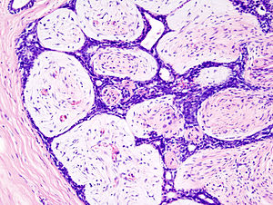
| 組織病理切片(苏木精-伊红染色) | |
| 症状 | 乳房腫塊 |
| 分类和外部资源 | |
| 醫學專科 | 婦科學 |
| ICD-11 | 2F30.5 |
| DiseasesDB | 1595 |
| MedlinePlus | 007216 |
| eMedicine | 345779 |
流行病學
编辑乳房纖維腺瘤最常出現於生育年齡的女性,特別是小於30歲,不過仍可生長於各種年齡層[5]。
症狀
编辑典型症狀為生育年齡的女性胸部出現單顆無痛結實、邊界清晰、生長緩慢、可自由推動的腫塊,通常小於3公分[4][6][7]。在少數情況,同側或兩個乳房中,同時或先後出現多顆腫塊,甚至可以成長到20公分;或者在乳房攝影中發現結節、鈣化或觸摸不出來的小型腫塊[5]。當腫瘤內出血時,疼痛可能是某些個案的第一個徵候[2]。
乳房纖維腺瘤大部分是良性的,癌化的機會在1000人中约有3人。因為乳房纖維腺瘤從小葉中生長出來,小葉癌是乳房纖維腺瘤裡面最常發現的癌症,也有期刊報告過乳腺管原位癌的發生[2]。
發生原因
编辑乳房纖維腺瘤與荷爾蒙有關,經常在停經後消退。大量攝取蔬菜水果、高生育數量、少使用口服避孕藥以及適當運動與腫瘤低發生率相關[9]。
病理學
编辑巨觀
编辑腫瘤呈現圓形或卵形,具有彈性與光滑表面,切面通常為同質紮實的灰白色或黃褐色組織。小管周型具有完整被膜與漩渦狀的外表,而小管內型則擁有不完整的被膜[10]。
微觀
编辑乳房纖維腺瘤呈現基質與上皮組織的雙重增生,可以呈現小管周型(基質圍繞著上皮結構)和小管內型(增生的基質壓縮上皮結構而形成裂縫)兩種型態,但沒有臨床表現的差別[4]。與惡性腫瘤相比,乳房纖維腺瘤擁有較少血管的基質[4][7][10]。而且,在單一終末腺管單元中,纖維母細胞基質包圍增生的上皮而形成管狀空間[11]。
分子
编辑診斷
编辑乳房纖維腺瘤的診斷方法包括:臨床檢查、醫學超音波檢查、乳房攝影術,和細針抽吸細胞診斷。
-
組織照(蘇木精-伊紅染色),小管周型(左下方)和小管內型(右上方)
-
細針抽吸細胞診斷(吉姆薩或迪夫快速染色)
-
細針抽吸細胞診斷(柏氏染色)
-
病理組織照,粗針切片(蘇木精-伊紅染色)
-
病理組織照(蘇木精-伊紅染色)
-
超音波照
治療
编辑大多數的乳房纖維腺瘤只需要持續追蹤,其他則是接受切除手術,只需要少量切口邊緣組織[10][14]。因為細針抽吸細胞診斷常是可靠的診斷工具,有些醫師決定不開刀切除,而使用臨床理學檢查與乳房攝影追蹤病灶的生長速度。小於50歲的女性每個月小於16%的生長速度,或大於50歲女性有小於13%的速度,是可以持續追蹤不需要開刀的安全速度[15]。
奧美昔芬對某些乳房纖維腺瘤患者有效[16]。
在完全切除後,乳房纖維腺瘤大部分沒有復發的現象[4]。
冷凍療法
编辑美國食品藥品監督管理局在2001年核准冷凍療法為手術切除外的一種安全有效、最小侵入性治療選項[17]。在此治療過程中,超音波導引探針進入乳房腫瘤部位,然後使用極度低溫來摧毀異常細胞[18],細胞隨後被重新吸收到體內。這項療法可以在門診進行,並只需使用局部麻醉,比起手術留下較少疤痕組織,而且較少乳房變形的後遺症[18]。
參見
编辑參考文獻
编辑- ^ Greenberg R, Skornick Y, Kaplan O. Management of breast fibroadenomas. J Gen Intern Med. 1998, 13 (9): 640–5.
- ^ 2.0 2.1 2.2 Samuel Pilnik (编). Common Breast Lesions: A Photographic Guide to Diagnosis and Treatment. Cambridge University Press. 2003: 4 [2018-09-06]. ISBN 9780521823579. (原始内容存档于2018-09-06). (页面存档备份,存于互联网档案馆)
- ^ 22-251c.Fibroadenomas at Merck Manual of Diagnosis and Therapy Home Edition
- ^ 4.0 4.1 4.2 4.3 4.4 Tavassoli, F.A.; Devilee, P. (编). World Health Organization Classification of Tumours: Pathology & Genetics: Tumours of the breast and female genital organs. Lyon: IARC Press. 2003. ISBN 92-832-2412-4.[页码请求]
- ^ 5.0 5.1 Lakhani, S.R.; Ellis. I.O. (编). WHO Classification of Tumours of the Breast. Lyon: IARC Press. 2012: 142 [2018-09-08]. ISBN 9789283224334. (原始内容存档于2018-01-12). (页面存档备份,存于互联网档案馆)
- ^ DeMay, M. Practical Principles of Cytopathology Revised. ASCP Press. : 2007. ISBN 0-89189-549-3.[页码请求]
- ^ 7.0 7.1 Pathology Outlines Website. [1] (页面存档备份,存于互联网档案馆) Accessed 12 February 2009.
- ^ Shin SJ, Rosen PP. Bilateral presentation of fibroadenoma with digital fibroma-like inclusions in the male breast. Archives of Pathology & Laboratory Medicine. July 2007, 131 (7): 1126–9. PMID 17617003. doi:10.1043/1543-2165(2007)131[1126:BPOFWD]2.0.CO;2 (不活跃 2017-08-21).
- ^ Nelson ZC, Ray RM, Wu C, Stalsberg H, Porter P, Lampe JW, Shannon J, Horner N, Li W, Wang W, Hu Y, Gao D, Thomas DB. Fruit and vegetable intakes are associated with lower risk of breast fibroadenomas in Chinese women. The Journal of Nutrition. July 2010, 140 (7): 1294–301. PMC 2884330 . PMID 20484549. doi:10.3945/jn.109.119719.
- ^ 10.0 10.1 10.2 Rosen, PP. Rosen's Breast Pathology 3rd. 2009. ISBN 978-0-7817-7137-5.[页码请求]
- ^ Fibroadenoma of the breast. [2007-12-15]. (原始内容存档于2009-03-10). (页面存档备份,存于互联网档案馆)
- ^ Lim WK, Ong CK, Tan J, Thike AA, Ng CC, Rajasegaran V, Myint SS, Nagarajan S, Nasir ND, McPherson JR, Cutcutache I, Poore G, Tay ST, Ooi WS, Tan VK, Hartman M, Ong KW, Tan BK, Rozen SG, Tan PH, Tan P, Teh BT. Exome sequencing identifies highly recurrent MED12 somatic mutations in breast fibroadenoma. Nature Genetics. August 2014, 46 (8): 877–80. PMID 25038752. doi:10.1038/ng.3037.
- ^ Piscuoglio S, Murray M, Fusco N, Marchiò C, Loo FL, Martelotto LG, Schultheis AM, Akram M, Weigelt B, Brogi E, Reis-Filho JS. MED12 somatic mutations in fibroadenomas and phyllodes tumours of the breast. Histopathology. November 2015, 67 (5): 719–29. PMC 4996373 . PMID 25855048. doi:10.1111/his.12712.
- ^ Rosai, J. Rosai and Ackerman's Surgical Pathology 9th. 2004. ISBN 978-0-323-01342-0.[页码请求]
- ^ Gordon PB, Gagnon FA, Lanzkowsky L. Solid breast masses diagnosed as fibroadenoma at fine-needle aspiration biopsy: acceptable rates of growth at long-term follow-up. Radiology. October 2003, 229 (1): 233–8. PMID 14519878. doi:10.1148/radiol.2291010282.
- ^ Dhar A, Srivastava A. Role of centchroman in regression of mastalgia and fibroadenoma. World Journal of Surgery. June 2007, 31 (6): 1178–84. PMID 17431715. doi:10.1007/s00268-007-9040-4.
- ^ Management of Fibroadenomas of the Breast (PDF). [2018-09-15]. (原始内容存档 (PDF)于2017-07-10). (页面存档备份,存于互联网档案馆)
- ^ 18.0 18.1 WebMD — Cryotherapy Shrinks Benign Breast Lumps. [2018-09-15]. (原始内容存档于2014-02-08). (页面存档备份,存于互联网档案馆)